

Sir Edward James Smith described three species of Pyrola in 1814 collected from West Redonda Island (BC, Canada) based on differences among these species in leaf morphology. Subsequently, taxonomists had trouble distinguishing among these taxa in other parts of the species’ ranges, largely because leaf size, shape, and coloration are highly variable, especially in P. picta and P. dentata, both of which have expanded laminae. Figure 1 (left) shows the stereotypical leaf morphology of each species.
Examining the micromorphology of leaf surfaces to see how stomatal density and arrangement, and epicuticular wax deposition (Fig. 2) vary among species across multiple climatic gradients provides more information about how members of the Pyrola picta species complex respond to climatic conditions. I have been examining leaves from populations across the Sierra Nevada to sample a broad latitudinal range and precipitation gradient (from arid east to the relatively mesic west side).

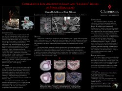
Anatomical differences in cell size and arrangement in leaves of each species also differ. Pyrola picta is an evergreen species, so secondary growth in the petioles and lamina of leaves older than one year is quite obvious. Leaves of different ages can be easily identified on the basis of coloration, texture, and position on the stem (Fig.3).

Serial sections of leaves reveal anatomical differences between leaves in P. picta and P. aphylla, including differences in the arrangement of vascular tissues (Fig. 4). I am using my findings to develop a model for leaf evolution in parasitic plants.
PREPARATION OF TISSUES FOR MICRO-MORPHOLOGICAL AND ANATOMICAL STUDY
 In order to study leaf, stem, root, or floral tissues in high detail, these tissues typically must be preserved so that the cell walls and contents don’t deteriorate.
